Back Bones Diagram / Anatomy Of The Spine Globus Medical / Lower jaw (mandible) collar back of skull (occipital bone).
Dapatkan link
Facebook
X
Pinterest
Email
Aplikasi Lainnya
Back Bones Diagram / Anatomy Of The Spine Globus Medical / Lower jaw (mandible) collar back of skull (occipital bone).. Master fuse box manitou wiring diagrams mazda fuse box marathon wiring schematics marine engine diagram mazda b2200 wiring maytag wiring diagram dryer map. These bones work together to provide flexibility to the trunk, support the muscles of the trunk, and protect the spinal cord and spinal nerves of the back. Fishbone diagram or ishikawa diagram is a modern quality management tool that explains the cause and effect relationship for any quality issue that has arisen or that may arise. We also discuss what are osteons, what are canaliculi. This shopping feature will continue to load items when the enter key is pressed.
Disk herniation and gout, sciatica and spinal stenosis, osteoporosis diagram with back nerves and bones pain. The cranial bones include occipital bone, two parietal bones, frontal bone, two temporal bones, sphenoid bone, and the ethmoid bone. Bone science human diagram anchor chart human body health back skeleton. The bones of the chest — namely the rib cage and spine — protect vital organs from injury, and also provide structural support for the body. Start learning with our skeleton diagrams, bone labeling exercises and skeletal system quizzes!
Spinal Cord Column Spinal Cord Injury Information Pages from www.sci-info-pages.com The bones of the chest — namely the rib cage and spine — protect vital organs from injury, and also provide structural support for the body. Pngtree offers bone diagram png and vector images, as well as transparant background bone diagram clipart images and psd files. Cheek bone (zygoma) upper jaw (maxilla). In this article, we look at the structure and function of this bone and the injuries that can affect it. Master fuse box manitou wiring diagrams mazda fuse box marathon wiring schematics marine engine diagram mazda b2200 wiring maytag wiring diagram dryer map. We also discuss what are osteons, what are canaliculi. The top and both sides of the head are formed by the paired. In this video we discuss the structure of bone tissue and the components of bones.
This framework consists of many individual bones and cartilages.
All the bones in the body can be described as long bones or flat bones are composed of two thin layers of compact bone that surround a layer of cancellous. This shopping feature will continue to load items when the enter key is pressed. Neck vertebrae (7) (cervical vertebrae). These bones work together to provide flexibility to the trunk, support the muscles of the trunk, and protect the spinal cord and spinal nerves of the back. Cheek bone (zygoma) upper jaw (maxilla). In order to navigate out of this carousel please use your heading shortcut key to navigate to the next or previous heading. The cranial bones include occipital bone, two parietal bones, frontal bone, two temporal bones, sphenoid bone, and the ethmoid bone. Download the free graphic resources in the form of png, eps. Bone science human diagram anchor chart human body health back skeleton. Right superficial lymphatic vessels of back. Master fuse box manitou wiring diagrams mazda fuse box marathon wiring schematics marine engine diagram mazda b2200 wiring maytag wiring diagram dryer map. The foot bones shown in this diagram are the talus, navicular, cuneiform, cuboid, metatarsals and calcaneus. There also are bands of fibrous connective tissue—the ligaments and a diagram of the human skeleton showing bone and cartilage.
Start learning with our skeleton diagrams, bone labeling exercises and skeletal system quizzes! Lower back of the head. It helps in brainstorming to identify possible causes of a problem and in sorting ideas into useful categories. There also are bands of fibrous connective tissue—the ligaments and a diagram of the human skeleton showing bone and cartilage. A bone is a rigid tissue that constitutes part of the vertebrate skeleton in animals.
Sci Injuries How They Occur Level Of Injury from www.latinaproject.com Back, bones and human spin diseases explanation vector. Continue scrolling to read more below. Start learning with our skeleton diagrams, bone labeling exercises and skeletal system quizzes! The temporal bone is one of the thickest bones in the skull. Human bones diagram 12 photos of the human bones diagram human anatomy diagram back view organs, human anatomy diagram diaphragm, human anatomy diagram of ear. Neck vertebrae (7) (cervical vertebrae). Right superficial lymphatic vessels of back. Disk herniation and gout, sciatica and spinal stenosis, osteoporosis diagram with back nerves and bones pain.
Bone science human diagram anchor chart human body health back skeleton.
It helps in brainstorming to identify possible causes of a problem and in sorting ideas into useful categories. Cheek bone (zygoma) upper jaw (maxilla). All the bones in the body can be described as long bones or flat bones are composed of two thin layers of compact bone that surround a layer of cancellous. The foot bones shown in this diagram are the talus, navicular, cuneiform, cuboid, metatarsals and calcaneus. Test your knowledge of the main bones of the body with our unlabeled diagram (download below). Start learning with our skeleton diagrams, bone labeling exercises and skeletal system quizzes! In this video we discuss the structure of bone tissue and the components of bones. The top and both sides of the head are formed by the paired. Fishbone diagram or ishikawa diagram is a modern quality management tool that explains the cause and effect relationship for any quality issue that has arisen or that may arise. Neck vertebrae (7) (cervical vertebrae). Back anatomy diagram lower bones rear view of human skeletal system showing upper back stock photo anatomy of the spine and back anatomy of the back bones sciences. Disk herniation and gout, sciatica and spinal stenosis, osteoporosis diagram with back nerves and bones pain. The cranial bones include occipital bone, two parietal bones, frontal bone, two temporal bones, sphenoid bone, and the ethmoid bone.
The temporal bone is one of the thickest bones in the skull. Back anatomy diagram lower bones rear view of human skeletal system showing upper back stock photo anatomy of the spine and back anatomy of the back bones sciences. Bones protect the various organs of the body, produce red and white blood cells, store minerals. Cheek bone (zygoma) upper jaw (maxilla). In this article, we look at the structure and function of this bone and the injuries that can affect it.
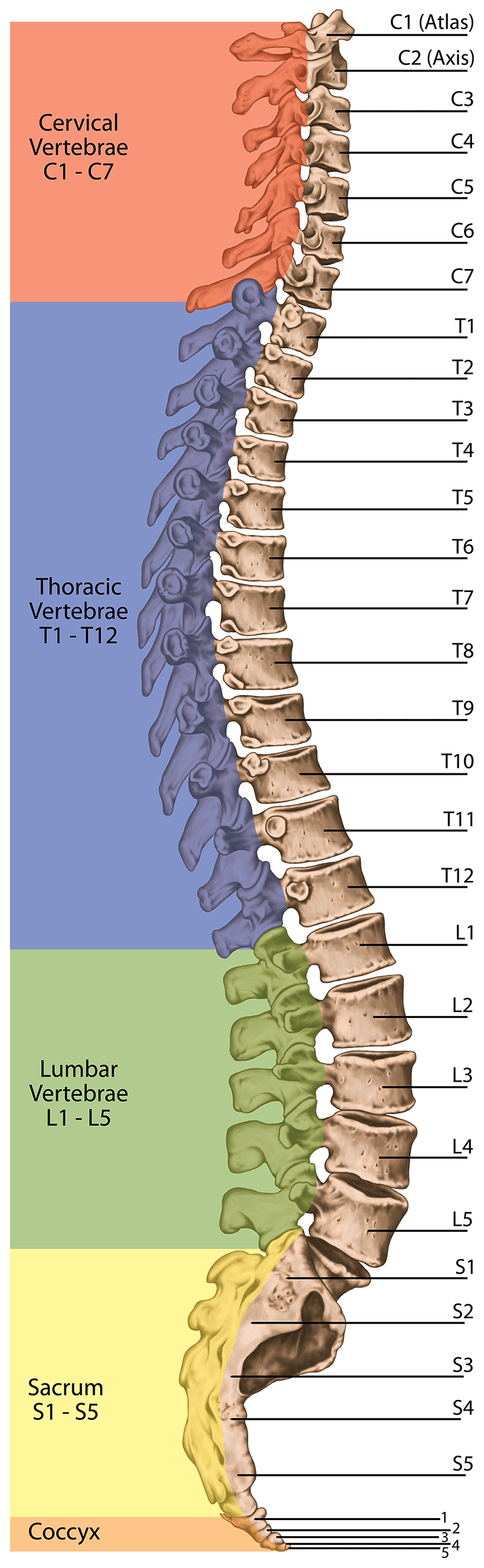
Spine Structure Function Parts Segments Spine Problems Spine Health from www.clevelandclinic.org Bone science human diagram anchor chart human body health back skeleton. The foot bones shown in this diagram are the talus, navicular, cuneiform, cuboid, metatarsals and calcaneus. This framework consists of many individual bones and cartilages. The top and both sides of the head are formed by the paired. Lower back of the head. The bones of the chest — namely the rib cage and spine — protect vital organs from injury, and also provide structural support for the body. In order to navigate out of this carousel please use your heading shortcut key to navigate to the next or previous heading. Test your knowledge of the main bones of the body with our unlabeled diagram (download below).
The bones of the chest — namely the rib cage and spine — protect vital organs from injury, and also provide structural support for the body.
Back anatomy diagram lower bones rear view of human skeletal system showing upper back stock photo anatomy of the spine and back anatomy of the back bones sciences. This shopping feature will continue to load items when the enter key is pressed. We also discuss what are osteons, what are canaliculi. Bones protect the various organs of the body, produce red and white blood cells, store minerals. Back, bones and human spin diseases explanation vector. Test your knowledge of the main bones of the body with our unlabeled diagram (download below). In order to navigate out of this carousel please use your heading shortcut key to navigate to the next or previous heading. Disk herniation and gout, sciatica and spinal stenosis, osteoporosis diagram with back nerves and bones pain. In this video we discuss the structure of bone tissue and the components of bones. Continue scrolling to read more below. The bones of the leg are the femur, tibia, fibula and patella. Start learning with our skeleton diagrams, bone labeling exercises and skeletal system quizzes! Human bones diagram 12 photos of the human bones diagram human anatomy diagram back view organs, human anatomy diagram diaphragm, human anatomy diagram of ear.

Komentar
Posting Komentar